Understanding Phlebitis and Infiltration
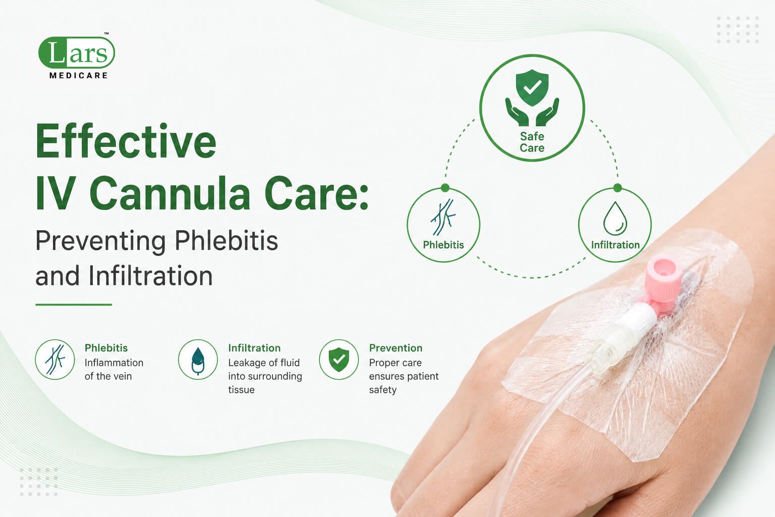
Phlebitis and infiltration are two critical complications associated with peripheral intravenous (IV) cannulation. Phlebitis, an inflammatory reaction of the vein, can occur due to mechanical irritation, chemical irritants from IV solutions, or infectious agents. This condition is characterized by localized redness, warmth, swelling, and pain along the vein where the IV cannula is inserted. Phlebitis can lead to serious complications, including the risk of thrombosis or systemic infection if left untreated.
In contrast, infiltration refers to the unintended leakage of IV fluids into the surrounding tissue instead of the bloodstream. This can result from a dislodged catheter or a vein that has become too small for the size of the IV cannula, leading to swelling, pain, and potential tissue damage. Symptoms of infiltration may include skin pallor, swelling at the infusion site, and a feeling of tightness in the affected area.
While both conditions can arise during IV therapy, their underlying causes and outcomes differ significantly. Phlebitis is predominantly caused by inflammation, whereas infiltration is a mechanical failure in IV delivery. Timely identification and management of these complications are essential for ensuring the comfort and safety of patients receiving IV therapy. Healthcare professionals should be educated about the signs and symptoms of both phlebitis and infiltration to take appropriate measures to mitigate risks. Adhering to best practices in IV cannula care, such as regular monitoring of the insertion site and proper catheter maintenance, can help prevent these complications.
Understanding phlebitis and infiltration is vital for healthcare practitioners involved in administering IV treatments. By recognizing the differences between these two conditions, along with their causes and potential complications, professionals can enhance patient safety and improve overall care outcomes during IV therapy.
Best Practices for IV Cannula Insertion
Inserting a peripheral IV cannula is a critical skill that requires meticulous attention to detail to ensure patient safety and minimize complications. The first step in this process is patient preparation. It is essential to explain the procedure to the patient, addressing any concerns they may have, which can reduce anxiety and promote cooperation. Assessing the patient’s medical history, including allergies and previous IV experiences, is also crucial to tailor the insertion technique to their specific needs.
The choice of site for IV insertion significantly impacts the success of the procedure. Optimal sites are typically located in the upper extremities, specifically the forearm or hand, as these areas tend to have larger veins that are less likely to cause discomfort and allow for easier infusion. Prioritize areas that are devoid of joints or areas with considerable flexion to prevent cannula dislodgement. Before proceeding, palpate the veins to ensure they are not only visible but also resilient, indicating good quality for cannulation.
Aseptic technique is paramount during the insertion process to prevent infection, including phlebitis and infiltration, which can lead to severe complications. Begin by washing hands thoroughly and utilizing appropriate personal protective equipment (PPE). Employ sterile drapes and cleanse the selected site with an appropriate antiseptic solution, allowing it to dry completely to enhance the effectiveness of the antimicrobial properties.
During the insertion itself, hold the cannula at the hub and insert it at a 15 to 30-degree angle to navigate smoothly through the skin and into the vein. Employ a gentle forward motion while simultaneously applying slight traction on the skin to stabilize the vein. Observing for a flashback of blood confirms successful intravascular access. With these best practices in place, healthcare professionals can reduce the risk of complications, thus promoting a safer and more effective IV cannulation experience for patients.
Ongoing Monitoring of IV Sites
Regular monitoring of intravenous (IV) sites is crucial in mitigating the risks of complications, particularly phlebitis and infiltration. By consistently assessing the IV sites, healthcare professionals can detect early signs of these conditions, thereby enhancing patient safety and comfort.
During the monitoring process, specific observations should be made. Clinicians should check for redness, warmth, swelling, pain, or tenderness around the IV insertion site, as these can indicate phlebitis, an inflammation of the vein. Additionally, signs of infiltration, which occurs when IV fluid leaks into surrounding tissues rather than entering the vein, include swelling, pallor, or a change in temperature at the site. Prompt recognition of these signs is imperative for timely intervention.
Different patient populations may warrant varying frequencies of assessment. For instance, critically ill patients or those with a history of phlebitis may require more frequent monitoring, perhaps every hour, to ensure any emerging issues are promptly addressed. Conversely, stable patients may be appropriately assessed every four to eight hours, depending on their condition and the type of IV therapy being administered.
It is also vital to educate patients about what to look for and encourage them to report any unusual sensations or changes at the IV site. This collaborative approach not only empowers patients but also contributes to a comprehensive monitoring strategy.
In conclusion, the ongoing evaluation of IV sites is an integral part of IV cannula care. Regular assessments enable healthcare providers to identify issues early on, thus reducing the complications associated with IV therapy and ensuring optimal patient outcomes.
IV Cannula Maintenance Techniques
Maintaining a peripheral IV cannula is crucial in preventing complications, such as phlebitis and infiltration. Daily care and regular maintenance routines help ensure the longevity and functionality of the cannula. A key component of this is the flushing protocol, which adequately clears the intravenous line of any residual medication or clotted blood. It is essential to follow the institution’s established protocols, usually recommending a saline flush before and after medication administration, as well as at regular intervals when the cannula is not in use.
Flushing not only maintains the patency of the IV line but also minimizes the risk of phlebitis, which is inflammation of the vein wall that can result from irritation caused by the cannula itself or the injected substances. Following the aseptic technique during flushing is imperative to reduce the risk of infection. Health professionals should ensure they are using appropriate syringe sizes and that the flush solution is free from contaminants.
Another vital aspect of IV cannula maintenance is the regular change of the dressing surrounding the site. Dressings should be inspected frequently for signs of drainage, bleeding, or other changes that could indicate complications. It is recommended to use transparent dressings where possible, as they allow for continuous monitoring of the insertion site. The frequency for changing dressings can vary based on organizational policy, often ranging from every 48 to 72 hours.
Lastly, implementing securement devices is essential to prevent movement and irritation of the cannula. Various types of securement devices such as adhesive tapes or specialized IV stabilization devices help keep the cannula in place, reducing the risk of accidental dislodgement or inadvertent trauma to the insertion site. Proper training on how to apply these devices effectively can greatly enhance patient comfort and safety.
Identifying Risk Factors
Phlebitis and infiltration are significant complications associated with intravenous (IV) cannulation. Understanding the risk factors that contribute to these conditions is crucial for implementing effective IV cannula care protocols. Risk factors can be broadly classified into individual and environmental categories.
Firstly, patient-related factors play a vital role in determining the likelihood of phlebitis and infiltration. Age is a prominent factor; older adults often exhibit fragile veins and decreased elasticity, increasing the risk for complications during and after cannulation. Furthermore, patients with comorbidities such as diabetes, obesity, or peripheral vascular disease may have compromised vascular integrity, further heightening their susceptibility.
Another important consideration is the type of medication administered through the IV cannula. Certain medications, particularly irritants or vesicants, can cause local tissue damage and inflammation, leading to phlebitis. For example, chemotherapeutic agents and hypertonic solutions require special attention during administration.
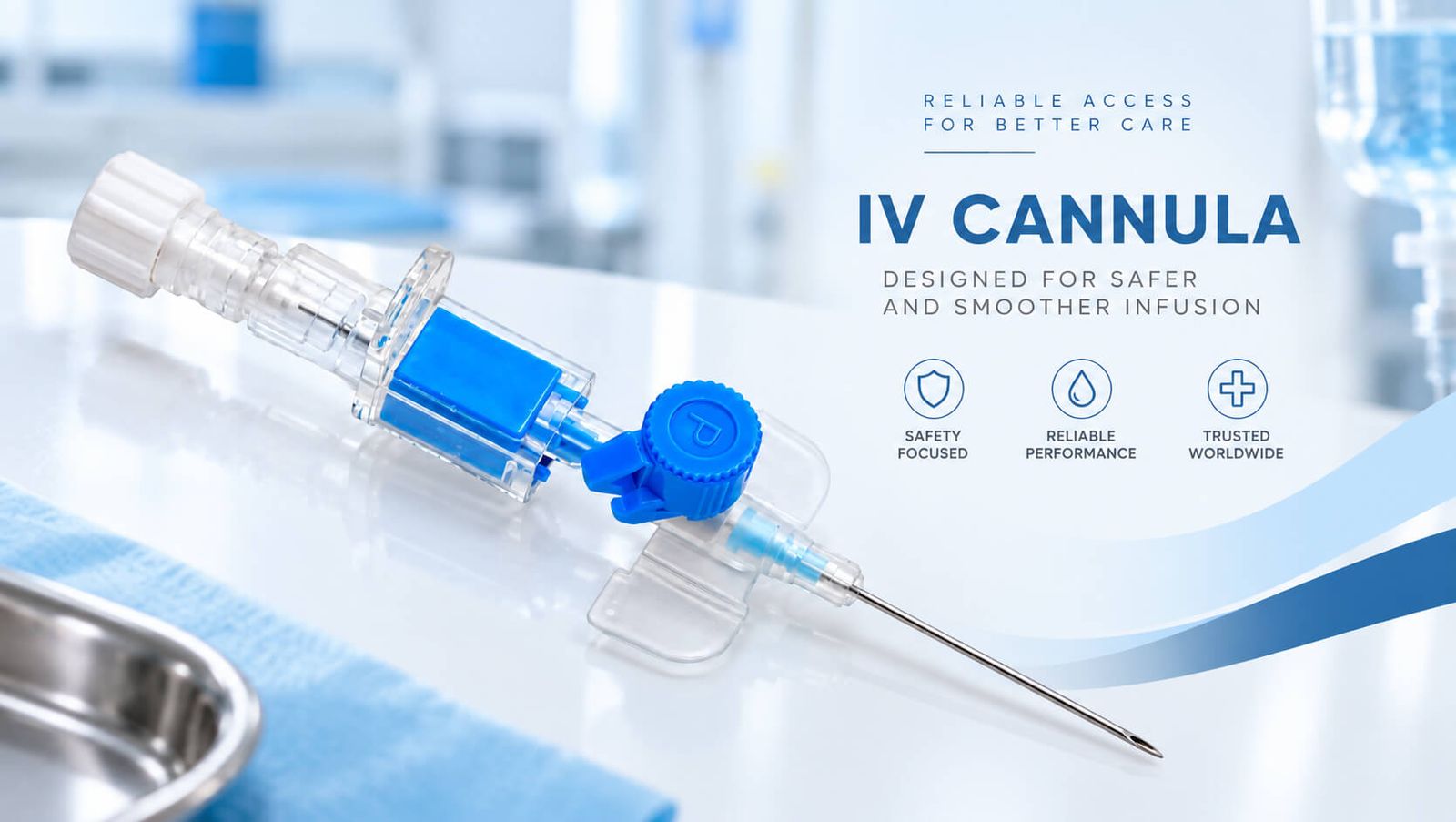
Additionally, the choice of cannula size can influence risk. Larger gauge cannulas may pose a greater risk of trauma to the vein wall compared to smaller-sized ones. Therefore, assessing the appropriate cannula size based on the patient’s vein condition and the therapy prescribed is essential for minimizing complications.
Environmental factors also contribute to the risk of developing phlebitis and infiltration. The availability of adequate training for healthcare professionals in IV cannulation techniques can impact the outcomes. Poor technique during insertion or maintenance of the IV line can lead to unnecessary patient suffering. Furthermore, the presence of a sterile environment is critical; improper aseptic technique can lead to infection, exacerbating venous inflammation.
In conclusion, both patient-related and environmental risk factors are essential considerations in preventing phlebitis and infiltration during IV therapy. By recognizing these risks and taking appropriate measures, healthcare providers can enhance patient outcomes and enhance the effectiveness of IV cannula care.
Education and Training for Healthcare Providers
The complexity of modern healthcare necessitates that healthcare providers maintain a high level of competency, particularly in areas such as peripheral intravenous (IV) cannula care. As phlebitis and infiltration are common complications associated with IV therapy, it is imperative that ongoing education and training programs are implemented to foster the skills required for effective IV management.
A well-structured training program can significantly enhance the proficiency of healthcare professionals in peripheral IV care. These programs should encompass both theoretical knowledge and practical skills, addressing the latest best practices in cannula insertion, maintenance, and removal. Moreover, education regarding the identification and management of potential complications will empower providers to deliver safer patient care.
Incorporating evidence-based research into these training modules is crucial. As new studies emerge, healthcare providers must adapt their practices accordingly to ensure they are employing the most effective techniques. Periodic refresher courses and workshops can facilitate this ongoing education, allowing practitioners to update their knowledge and skills regularly.
Another critical aspect of training involves the application of simulation-based learning. Hands-on practice in a controlled environment allows providers to refine their techniques and boost confidence prior to real-world application. Furthermore, mentorship programs where experienced practitioners guide less experienced staff can cultivate an environment of continuous improvement and knowledge sharing.
Ultimately, the goal of implementing robust education and training programs is to minimize the risks associated with peripheral IV therapy, specifically by preventing complications such as phlebitis and infiltration. By prioritizing optimal training standards and staying informed about advancements in the field, healthcare professionals will be better equipped to ensure high-quality patient care.
Patient Education on IV Care
Patient education plays a crucial role in the successful management of intravenous (IV) therapies. When patients understand their treatment process, including the purpose of the IV cannula, they are better equipped to participate actively in their own care. Education should begin as soon as the decision is made to initiate IV therapy. Healthcare professionals should communicate clearly about what patients can expect during the procedure, including the placement of the cannula and the sensations associated with it.
During the education process, it is essential to inform patients about the signs and symptoms of potential complications, such as phlebitis and infiltration. Patients should be instructed to watch for signs like swelling, redness, or tenderness at the IV site. They must also be aware of systemic reactions like fever or chills, which may indicate an infection. By recognizing these signs early, patients can report concerns promptly, leading to timely medical intervention.
Moreover, it is imperative to emphasize the importance of adhering to care instructions provided by healthcare professionals. Patients should understand that proper care and compliance can significantly reduce the risk of complications associated with IV therapy. This includes instructions on how to maintain the cannula site, keep it clean, and what activities to avoid during treatment. Strategies such as providing pamphlets, utilizing teach-back methods, and engaging family members can enhance patient understanding and retention of this vital information.
In addition, healthcare providers may wish to tailor educational materials to individual patient needs by considering cultural backgrounds and literacy levels. By employing various teaching methods and ensuring that patients feel comfortable asking questions, healthcare professionals can foster an informative and encouraging environment. Ultimately, effective patient education on IV care empowers patients to take an active role in their treatment and enhances overall safety and efficacy in their IV therapy journey.
Responding to Complications
Recognizing and effectively responding to complications associated with IV cannula use, particularly phlebitis and infiltration, is crucial for maintaining patient safety and comfort. Healthcare professionals must be vigilant in monitoring the IV site for any signs that may indicate these complications. Common indicators of phlebitis include redness, swelling, and warmth at the site, along with pain or tenderness upon palpation. Infiltration, on the other hand, may present as swelling in the area surrounding the cannula, along with possible changes in skin color and an increase in local tissue tension.
Upon observing any indications of phlebitis or infiltration, the first step in management is to assess the severity of the complication. In instances where phlebitis is mild, it may be appropriate to simply discontinue the IV cannula while monitoring the patient closely. Conversely, if signs of infiltration are present, the healthcare provider should take immediate action to remove the IV cannula to prevent further tissue damage.
In both cases, appropriate documentation of the findings is essential. This includes noting the date and time of the assessment, specific observations related to phlebitis or infiltration, and any actions taken, such as the removal of the IV cannula. Additionally, it is important to document any treatments administered to mitigate discomfort, such as cold compresses for swelling or medical interventions in cases of more severe phlebitis.
Moreover, following the removal of the cannula, clinicians should assess the need for replacement IV access, either at the same site if the tissue condition allows or at a different site altogether. Providing patient education about what signs and symptoms to watch for post-removal is also beneficial as it empowers patients to report any issues promptly, ensuring timely intervention.
Future Perspectives in IV Therapy
The field of intravenous (IV) therapy is increasingly evolving, with significant advancements aimed at enhancing patient safety and optimizing the efficacy of IV cannula care. One of the most notable trends involves the development of innovative materials for IV cannula devices. These materials are designed to reduce the risk of complications such as phlebitis and infiltration. For instance, new generation cannulas are being manufactured using advanced biocompatible polymers that can reduce irritation and the potential for inflammation at the insertion site.
Additionally, technology integration plays a crucial role in the future perspectives of IV therapy. Smart IV devices equipped with sensors and connectivity features are gaining ground, allowing healthcare providers to monitor patients more effectively. These devices can provide real-time data on cannula patency and alert the medical team to any complications such as infiltration or occlusion. This proactive approach significantly enhances the management of IV therapies while ensuring that patient comfort and safety are prioritized.
Furthermore, there is a growing emphasis on training and education to improve overall IV cannula care. With the introduction of new technologies and materials, healthcare professionals must stay informed about the best practices and guidelines. Simulation-based training and continuing education programs are becoming integral components of nursing curricula, ensuring that practitioners are well equipped to handle the latest advancements in IV therapy.
Overall, the future of IV therapy is geared towards improving patient outcomes through innovative solutions. As research continues to unveil more effective materials and technologies, the goal remains clear: to deliver IV care that minimizes risks and maximizes efficiency. By adopting these emerging trends, healthcare providers can significantly enhance the quality of IV cannula care, ultimately leading to better patient satisfaction and reduced healthcare costs.